The explosions of atomic bombs in Hiroshima and Nagasaki and the accident in Chernobyl clearly showed the serious health consequences of ionizing radiation.
Consequences of exposure to ionizing radiation can appear immediately when the threshold value is exceeded (acute radiation syndrome) or can be delayed and occur with the gradual accumulation of radiation doses over time (carcinogenesis).
per 100 mSv Because equal radiation exposure can cause cancer-related death in 1/100,000 patients, the International Atomic Energy Association (IAEA) has set the following limits:
▶ 50 mSv per year for total body exposure (or 20 mSv annually for 5 years)
▶ 500 mSv for single organ irradiation
Suffering from urolithiasis radiation exposure during examination of patients:
☑ Standard CT 10 mSv,
☑ Low-dose CT ~3 mSv,
☑ General radiography 0.7 mSv
☑ urography 2.1-3 mSv
☑ Ultrasound - 0 mSv
How to determine the received radiation dose?
Each modern X-ray machine or computer tomography has a built-in dosimeter and displays the number of mSv received immediately after the study. This indicator should be recorded in the study protocol.
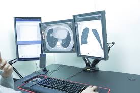
What is the best way to identify stones?
The "gold standard" for the diagnosis of ICD is low-dose tomography (CT)
Its sensitivity is about 93.1% and specificity is about 96.6%.
The sensitivity and specificity of X-ray and ultrasound are much lower (respectively 44-77% and 45-94%) :)
The sensitivity of ultrasound and X-rays is even lower for small stones:
⏺ Ultrasound examination does not poorly show stones smaller than 3 mm, because such particles may not create an acoustic shadow, and the doctor must differentiate between echogenic renal sinus fat and stone particles.
⏺ Ultrasound overestimates stone size in 50% of cases compared to CT
⏺ X-rays underestimate (underestimate) stone size.
Therefore, low-dose CT remains the only way to evaluate small stones:
What is low-dose renal CT?
This is a scanning mode aimed at detecting stones in the urinary system. Because stones have very different x-ray properties than body tissues, lower-powered scanners have become possible.
With a low-dose CT scan, the radiologist only images the presence of stones and not other structures.
Low-dose CT uses 30 milliamperes (mA) of current instead of the 100 mA of standard protocols.
The scan takes only 10-15 seconds.
All of this results in reduced radiation exposure without loss of sensitivity or specificity in the diagnosis of urolithiasis.
The main disadvantages of low-dose CT protocols are body mass index > 30 kg / m2 is considered a decrease in sensitivity.
Normal BMI from 18.5 to 25
A person with a BMI from 25 to 30 is overweight (before obesity)
Obese people have a BMI > will be 30 kg/m2.
To calculate the body mass index, you need to divide your body weight in kilograms by the square of your height in meters.
For example, a person weighs 77 kg and is 170 cm tall.
Therefore, in this case, BMI is equal to: BMI = 77: (1.70 × 1.70) ≈ 26.64 kg / m².
So, the official According to the standard protocol, a patient with stone formation can undergo 15 low-dose CT scans or 5 CT scans per year.
The radiation dose received by the patient (and the doctor) during surgery should not be neglected❗
In addition, the results of surveys of foreign endourologists show that the level of knowledge and understanding of mechanisms, harm and safe limits is not clear. (Lazaros Tzelves, Knowledge of radiation exposure and adherence to protective equipment: where are the endourologists? ESUT/EULIS survey, 2020).